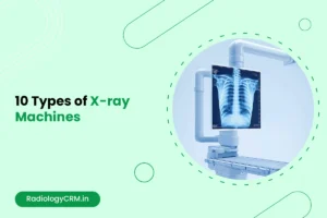
X-ray machines have revolutionized the way we diagnose diseases, monitor health conditions, and even inspect materials. Since the discovery of X-rays in 1895 by Wilhelm Roentgen, these machines have evolved significantly, leading to a variety of types that serve different purposes across various industries. From medical applications to security checks, X-ray machines are indispensable tools.
Whether you run a small clinic, regional hospital, specialized imaging center, or have started your own radiology equipment manufacturing company understanding these machine types is very important. It helps you make smarter equipment investments and business decisions.
In this blog post, we’ll dive deep into 10 types of X-ray machines, their uses, and how they work.
What are X-Ray Machines?
X-ray machines are medical devices that generate high-energy electromagnetic radiation to create images of internal body structures. These machines produce invisible energy beams with wavelengths ranging from 10 nanometers to 10 picometers, allowing them to penetrate human tissue and create diagnostic images on digital detectors or film.
X-ray machines operate through a simple yet sophisticated process. The machine contains an X-ray tube with a tungsten cathode (filament) that gets heated by electrical current. This heating releases electrons through thermionic emission, which are then accelerated at high speed toward a positively charged tungsten anode using electrical potential between 50-150 kilovolts.
When these high-energy electrons strike the anode target, approximately 1% of their energy converts into X-ray photons, while 99% becomes heat. The resulting X-rays travel in all directions from the focal spot, but collimators focus them into a controlled beam directed at the patient.
Different body tissues absorb X-ray energy at varying rates based on their density and atomic composition. Dense structures like bones absorb more X-rays and appear white on images, while soft tissues like muscles, organs, and blood allow more X-rays to pass through and appear darker gray.
This differential absorption creates contrast that allows healthcare professionals to visualize internal structures without invasive procedures. The remaining X-rays that pass through the body reach the image detector, where they create the final diagnostic image.
Boost your radiology business efficiency with RadiologyCRM.
Automate your quotes, contracts, leads, and billing—manage everything in one place.

What Makes Each X-ray Machine Type Different?
Before we explore specific machine types, you need to understand the core differences. X-ray machines vary by:
- Power output (measured in kV and mA)
- Image capture method (digital vs. film-based)
- Mobility (fixed, mobile, or portable)
- Specialized applications (general radiography vs. specific body regions)
- Real-time vs. static imaging capabilities
These factors directly impact image quality, patient throughput, radiation exposure, and total cost of ownership.
List of Different X-Ray Machines Types
1. Conventional (Fixed) X-ray Machine: The Standard Imaging Tool
The conventional X-ray machine is the most familiar type. This classic machine uses a single beam of X-rays directed at a part of the body, which then passes through to produce an image on a film or a digital detector. The resulting image is a 2D representation of the internal structure.
Applications:
- Used extensively to detect fractures, joint dislocations, and abnormalities in bones.
- Often used for chest imaging to diagnose pneumonia, lung infections, and lung cancer.
- Also employed in dental X-rays to assess tooth decay and bone density.
- High-volume hospitals and imaging centers
- Teaching institutions requiring diverse imaging capabilities
- Facilities performing complex radiographic procedures
- Emergency departments with 24/7 operations
Why It Matters:
Conventional X-rays are fast, inexpensive, and provide a clear, basic view of bone structures and certain soft tissues. They are the go-to for quick assessments in emergency rooms and doctor’s offices.
2. Computed Tomography (CT) Scanner: For Detailed, Cross-sectional Images
While conventional X-rays offer a 2D view, CT scanners provide detailed 3D cross-sectional images of the body. These machines use multiple X-ray images taken from various angles around the body, which are then processed by a computer to create detailed images of organs, blood vessels, and tissues.
Slice technology:
- Single-slice: Basic CT imaging
- Multi-slice: 16, 32, 64, 128+ detector rows
- Dual-source: Two X-ray tubes for cardiac imaging
CT scanners demand massive electrical capacity (100-120 kW) and specialized cooling systems due to heat generation from continuous X-ray production.
Applications:
- Ideal for detecting and diagnosing cancers, cardiovascular diseases, and neurological conditions.
- Crucial in trauma care, especially when a comprehensive view of internal injuries is needed.
- CT scans are frequently used to assess soft tissues, bones, and even blood vessels for conditions like stroke or tumors.
Why It Matters:
CT scans provide a much more detailed view compared to conventional X-rays, making them invaluable in complex diagnoses. Their ability to show cross-sections of the body helps doctors to spot hidden problems that traditional X-rays might miss.
3. Mammography Machine: Detecting Breast Cancer Early
Mammography is a specialized X-ray technique designed specifically for imaging the breast tissue. This machine uses low-dose X-rays to create detailed images of the breasts and detect early signs of cancer or other abnormalities.
Specialized components:
- Molybdenum or tungsten targets
- Beryllium windows for low-energy X-ray transmission
- Automatic exposure control designed for breast tissue
- Anti-scatter grids optimized for mammography
Applications:
- Primarily used to screen for breast cancer in women, especially those over the age of 40 or with a family history of the disease.
- Mammography can help detect tumors, cysts, and other breast conditions before they become palpable.
Why It Matters:
Early detection of breast cancer is crucial for effective treatment. Mammography machines are the primary tool used to catch cancer at its earliest and most treatable stages.
4. Fluoroscopy Machine: Real-Time Imaging for Medical Procedures
Unlike static X-ray images, fluoroscopy provides real-time video X-ray images of the body in motion. This type of imaging is used to observe internal organs or guide medical procedures that require real-time imaging.
Applications:
- Frequently used during diagnostic procedures like barium swallows or gastrointestinal studies.
- Used to guide medical professionals during catheter insertions, joint injections, and even some surgeries, allowing them to see the live positioning of tools.
Why It Matters:
Fluoroscopy provides dynamic, continuous imaging that allows healthcare providers to see how the body is functioning during specific movements or treatments. It’s an invaluable tool in both diagnostics and interventional procedures.
Fluoroscopy involves higher radiation doses due to continuous imaging. Proper technique and dose monitoring are essential for patient and staff safety.
Hence, fluoroscopy rooms need specialized lead shielding, emergency power systems, and climate control for sensitive electronics.
5. Dental X-ray Machine: Precision for Oral Health
Dental X-rays are essential for monitoring the health of teeth and gums. These X-ray machines are smaller and specifically designed to focus on the mouth and jaw, providing detailed images of teeth, bones, and surrounding structures.
Dental X-rays use extremely low doses but require proper collimation, filtration, and positioning to minimize patient exposure.
Intraoral systems:
- Wall-mounted or mobile tube heads
- Digital sensors or phosphor plates
- Exposure times: 0.1-2.0 seconds
- Patient positioning aids
Applications:
- For detecting cavities, tooth decay, abscesses or bone loss.
- Assists with dental treatments, such as implants, braces or root canals.
- Can also spot diseases like oral cancer or infections in the jawbone.
Why It Matters:
Dental X-rays play a valuable role in oral health care, helping to prevent problems and diagnose issues that are not clinically visible. They enable the dentist to detect problems early and plan proper treatments.
6. Portable X-ray Machine: Bringing Imaging to the Patient
Portable X-ray machines are both small and mobile, for use in a variety of situations where easy movement is beneficial, especially when the intended location is an area where patients can’t easily go for X-Ray examination. They are frequently operated in emergency, intensive care or home visits for bedridden patients.
Size and weight advantages:
- Weight: 60-120 pounds
- Power output: 70-110 kV, 15-35 mA
- Battery operation: 8-12 hours continuous use
Applications:
- Commonly used in emergency departments and for patients who are unable to move to an imaging room.
- Can be used for quick assessments of broken bones, lung issues, or abdominal pain.
Why It Matters:
Portable X-rays are flexible and convenient, allowing physicians to obtain vital diagnostic information without requiring patients to go out of their room or bed. This can be absolutely critical, especially in emergency or life and death situations.
7. Dual-Energy X-ray Absorptiometry (DEXA): Assessing Bone Health
DEXA scans use X-rays at two different energy levels to measure bone mineral density. It is the most common method used to diagnose osteoporosis and assess fracture risk.
Applications:
- Used to evaluate bone density and assess conditions like osteoporosis, especially in postmenopausal women or elderly individuals.
- Helps doctors determine the risk of fractures and guide treatment plans for bone health.
Why It Matters:
DEXA is the standard for measuring bone health, which helps health care providers predict and prevent fractures caused by osteoporosis. It’s an easy, minimally invasive test that is crucial for monitoring bone diseases.
8. Radiographic Fluoroscopy (C-Arm): Real-Time Imaging in Surgery
C-Arm is a special type of fluoroscopy machine, designed like the letter “C” which provides real-time X-ray imaging during various medical procedures including surgeries. It rotates around the patient to take pictures of the area being treated from different angles.
Applications:
- Commonly used in orthopedic surgeries, spinal surgeries, and interventional procedures like angioplasty.
- Provides live imaging to guide surgeons in precise procedures, helping to avoid complications.
Why It Matters:
C-Arm is necessary for minimally invasive surgeries as it limits large incisions. Real-time imaging can assist surgeons in making accurate decisions during operations, leading to better patient outcomes.
9. X-ray Backscatter Scanner: Security Applications
X-ray backscatter scanners are instruments that can reveal the contents of a container, suitcase or other object using X-rays. Unlike the X-ray machines that direct slice through an object, these scanners find the backscatter from energy bouncing off its surfaces.
Applications:
- Primarily used in security settings, such as airport security or customs inspections, to detect contraband or explosives.
- Works in cargo inspections, detecting concealed threats without disrupting the package.
Why It Matters:
Backscatter X-ray scanners increase security by offering a non-invasive method for screening the contents of bags, vehicles and ships. They are useful when it comes to stop smuggling or conveyance of dangerous substances.
10. Bone Densitometer: Specialized Bone Health Imaging
A bone densitometer is a special X-ray test for measuring the density of the bones. This machine measures how much calcium and other minerals are in your bones by using low-level X-rays.
Applications:
- Primarily used to diagnose osteoporosis and assess the risk of fractures.
- Helps in monitoring the effectiveness of osteoporosis treatments and tracking bone health over time.
Why It Matters:
This device is crucial for detecting bone loss early, which is important in preventing fractures and managing conditions like osteoporosis. It’s a key tool in managing long-term bone health.
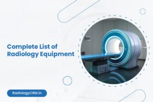
Here is the complete list of radiology equipment you should check.
Which X-ray Machine Type Fits Your Facility’s Needs?
Selecting the right X-ray equipment depends on several critical factors:
- Patient volume analysis: High-volume facilities will take advantage from the fixed DR system despite initial high cost. Lower-volume practices may find it more cost-effective to have CR or traditional systems.
- Service mix considerations: Emergency departments need mobile capabilities, while outpatient clinics may prioritize cost-effective general radiography systems.
- Space limitations: Portable or mobile units are more likely suitable options in a small facilities, to avoid dedicated rooms and infrastructure needed for fixed solutions.
- Budget constraints: Consider total cost of ownership including maintenance, training, and operational expenses beyond initial purchase price.
- Future growth planning: Invest in systems that can accommodate patient volume increases and additional service lines.
What Maintenance and Quality Assurance Do X-ray Machines Require?
- Daily quality checks: Technologists perform basic functionality tests, image quality assessments, and safety system verifications before patient use.
- Preventive maintenance: Manufacturers recommend quarterly or semi-annual service visits for calibration, component inspection, and software updates.
- Regulatory compliance: State and federal regulations require annual inspections by qualified medical physicists for radiation safety and image quality standards.
- Service contract considerations: Full-service contracts cost 8-12% of equipment value annually but provide predictable expenses and guaranteed response times.
Conclusion
The field of X-ray equipment is changing rapidly with technological advancements. Keeping up with new technology can help you make smart choices about equipment that will meet your facility’s needs for years to come.
Your X-ray equipment choices directly impact patient care quality, operational efficiency, and financial performance. Take time to thoroughly evaluate your specific needs, consult with equipment specialists, and consider long-term operational implications before making these significant investments.
Ready to upgrade your imaging capabilities? Start by assessing your current patient volume, service mix, and growth projections to identify which X-ray machine types will deliver the best return on your investment.