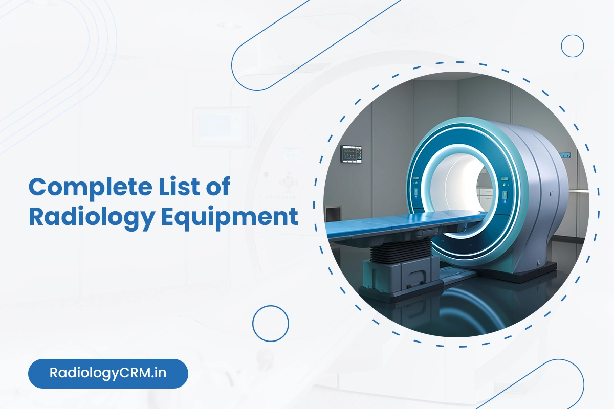
Radiology departments rely on a range of advanced equipment and systems to deliver accurate diagnoses and effective treatments.
In 2025, the global medical imaging market is projected to exceed $45 billion. It shows steady growth driven by rising demand for early disease detection, minimally invasive procedures, and improved patient outcomes.
Hospitals and diagnostic centers are investing in both established and emerging technologies to meet the needs of a growing and aging population.
Hence, understanding the full spectrum of radiology equipment is essential for healthcare leaders, radiologists, and technologists.
Whether you are preparing to upgrade your current radiology unit, forge a new facility, or just want to have an idea of the new products, then this guide will get into the nuts and bolts of every kind of radiology equipment, its functions, and the technology that have been released lately.
It covers diagnostic imaging machines, therapeutic radiation devices, and the supporting digital infrastructure.
If you are a seller of these equipment or systems, then have a look at RadiologyCRM, a CRM software specifically designed for radiology equipment sellers, vendors, distributors, retailers, etc.
Let’s get into more details –
Diagnostic Radiology Equipment (Imaging Systems)
1) X-ray Machine
X-ray technology remains the foundation of diagnostic imaging, with significant advancements enhancing its capabilities in coming years.
Modern X-ray machines now feature improved resolution and image quality while minimizing radiation exposure to patients.
The newest generation of X-ray systems feature artificial intelligence (AI) algorithms which quickly and accurately review images to assist radiologists in making better diagnoses. These A.I. systems can also spot abnormalities and early-stage diseases that may be difficult to pick out with a human eye.
Key advancements in X-ray technology include:
- High-Definition Imaging: Higher-resolution images are clearer for visualization and diagnosing.
- Lower Radiation Exposure: Developments in dose reduction prevent patients and X-ray technicians from unnecessary exposure.
- AI Integration: ML helps to detect the abnormalities and assist in diagnosis.
- Portable Options: Advancements in miniaturization have led to more effective portable X-ray devices.
The GE AMX 4 Plus and Siemens Mobilett Mira Max are among the top models, offering excellent balance between technology and cost. With 35kW generators coming standard, these machines provide more power than many competing options.
2) Portable X-ray Unit
Portable X-ray devices have made great advancements, and are necessary in health-care environments where patients are unable to move. Rapid diagnostic capabilities right at the patients’ bedside in emergency rooms, ICUs, doctor’s offices, ambulances or home health care settings, are critical for the early detection and quick treatment of disease.
The Carestream DRX-Revolution and the recently launched DRX-Rise portable leading the market with advanced functionality and features.
DRX-Revolution has a range of detector choices – DRX-1, DRX-plus, Lux detector – enabling different functionality to meet unique clinical requirements.
Siemens has recently upgraded their Elara Max with the new design, bigger high-contrast display, and incredibly wonderful Max detector which is capable of connection to a few systems. This interoperable performance is a new trend in portable X-ray technology and supports flexible workflow in busy medical environments.
- Modern portable X-ray units offer: Enhanced Mobility, lighter weight and improved maneuverability make these units easier to transport between patient rooms.
- Better Battery Life: Extended operation time allows for more examinations between charges.
- Wireless Connectivity: Immediate image transfer to PACS systems speeds up diagnosis and treatment decisions.
- Improved Image Quality: Despite their portable nature, these units now produce images comparable to fixed installations.
3) Digital Radiography (DR) System
Digital Radiography has changed medical imaging, providing a host of benefits over conventional film X-Rays.
DR systems are developing at a fast pace with better image quality, efficiency of workflow and patient care.
Key of the DR innovation is the improvements in detector technology. Newer detectors provide better resolution, allowing for more finely detailed images of anatomical structures that can contribute to more accurate patient diagnosis with less need to return for additional imaging.
Greater sensitivity means less radiation exposure for patients.
Key developments in DR systems include:
- Dynamic Range Expansion: Detectors that can capture a wider range of X-ray intensity levels offer both bony structures and soft tissue to be seen on a single image.
- Flat-Panel Detector Advancements: Flat-Panel detectors are now ubiquitous because of their lightweight and rugged characteristics combined with the relatively higher quality of the images, with faster acquisition times and improved image uniformity.
- AI Integration: AI-based algorithms help to improve the imaging quality, reduce noise and improve the contrast for the clearer visualization of tiny details.
- Wireless and Portable Systems: The increasing need for wireless and portable DR systems has enhanced workflow efficiency, especially in the point-of-care and emergency departments.
The utilization of AI in DR systems is among the most interesting new developments, and can include image reconstruction and highlighting, as well as computer-aided detection of abnormalities.
3) Fluoroscopy System
Fluoroscopy has evolved from a simple non-invasive imaging method to a sophisticated technology with advanced 3D capabilities.
Modern fluoroscopy systems provide real-time X-ray imaging that allows physicians to observe dynamic bodily functions and guide interventional procedures.
The latest digital fluoroscopy systems produce clearer, more detailed images than ever before. The improved image quality is particularly beneficial in guiding therapeutic procedures and surgeries, allowing for greater precision and better outcomes.
Key advancements in fluoroscopy technology include:
- Dose Reduction Technologies: Modern fluoroscopic machines have built-in functions that minimize exposure to radiation for both patients and staff while maintaining optimal image quality.
- Real-time Image Enhancement: Newer models provide improved visualization, enabling doctors to view minute details during procedures.
- 3D Fluoroscopy: 3D visualisation enhances navigation and spacial comprehension in complex interventions.
- Dynamic Digital Radiography (DDR): DDR records a series of X-ray images in quick succession so that doctors can see motion happen in real time, including lung function, joint movement, and swallowing issues.
Modern fluoroscopy systems create the perception of real-time imaging by capturing and displaying images at high frame rates, typically 25 or 30 frames per second. At these rates, motion appears continuous without visible flicker, allowing physicians to observe dynamic processes as they occur.
4) C-arm Machine (Mobile Fluoroscopy Unit)
C-arm machines have become essential tools in modern surgical settings, offering real-time imaging capabilities during procedures.
Named for their distinctive C-shaped arm that connects the X-ray source on one end and the detector on the other, these devices provide flexibility and precision during complex interventions.
These mobile fluoroscopy units are widely used during orthopedic, urological, gastroenterological, and cardiac procedures, as well as in pain management and emergency settings.
Key features of modern C-arm machines include:
- Enhanced Mobility: To obtain X-ray from different angles, the C-shaped arm can be moved horizontally, vertically, even its swivel axis can circle.
- Real-time Imaging: A fluoroscopy unit takes high-resolution X-ray images in real-time, and allows surgeons to perform a more precise surgery.
- Versatility: C-arms are adaptable to both fluoroscopy and radiography, thus acting like an all-purpose device in the surgical applications.
- Improved Workflow: Advanced systems offer streamlined controls and settings that enhance operational efficiency during procedures.
C-arm equipment flexibility makes it possible for surgeons to take images of the whole patient’s body from whichever angle is needed, supplying important visual support during minimally invasive treatments.
5) Angiography Machine
Angiography machines are specialized medical imaging systems used to visualize blood vessels, arteries, and veins in real-time. These sophisticated devices play a crucial role in diagnosing and treating various cardiovascular and neurological conditions.
The global angiography equipment market reached $11.3 billion in 2024 and is expected to grow at a CAGR of 6.0% to reach $19.0 billion by 2033. This growth is driven by the increasing prevalence of cardiovascular diseases, technological advancements, and the rising adoption of minimally invasive procedures.
Modern angiography systems incorporate several advanced features:
- AI-based Imaging: Integration of artificial intelligence enhances diagnostic accuracy and efficiency.
- 3D Angiography: Revolutionary visualization of blood vessels in three dimensions improves diagnostic precision.
- Reduced Radiation Exposure: Advanced algorithms and hardware improvements minimize radiation dose while maintaining image quality.
- Hybrid Systems: Combination of angiography with other imaging modalities like CT or MRI provides comprehensive diagnostic information.
The angiography systems segment dominates the market with a share of 22.1% in 2024, while X-ray technology leads the technology segment with a 40.9% market share.
Among procedures, intra-coronary angiography holds the largest share at 28.6%, reflecting its critical importance in cardiac care.
6) CT Scanner (Computed Tomography)
CT scanning technology has undergone remarkable transformations with innovations that redefine the traditional limits of these systems.
The biggest development in CT technology is the widespread adoption of photon-counting CT, which was first introduced by Siemens Healthineers with the NAEOTOM Alpha.
Photon-counting CT represents a complete rethink of how the underlying technology works. Unlike conventional CT scanners that measure X-ray energy in broad ranges, photon-counting detectors capture individual X-ray photons, leading to crisper images, lower radiation doses, and more accurate diagnoses.
Key advancements in CT technology include:
- Photon-counting Detectors: These detectors overcome the limits of traditional CT by streamlining what has always been a two-step process, resulting in superior image quality.
- Spectral CT Imaging: This technology enhances the ability to distinguish between different tissue types, particularly beneficial for early cancer detection and differentiating between benign and malignant growths.
- AI-enhanced Image Reconstruction: AI enhances CT scans by lowering noise and artifacts that may sometimes camouflage crucial diagnostic information.
- Reduced Radiation Exposure: Modern CT scanners achieve high-quality images with significantly lower radiation doses, addressing long-standing concerns about radiation exposure.
In cardiology, Coronary computed tomography angiography (CCTA) is increasingly being used as the first-line test in the evaluation of chest pain.
7) MRI Scanner (Magnetic Resonance Imaging)
Over the past decades, MRI has gained tremendous importance in diagnostic imaging with substantial technical advances expanding the its role in neurological, musculoskeletal, and cardiovascular studies.
There are a number of enhancements including technology advancements that allow for advanced ultrafast imaging with up to 50% reduction in scan time with excellent image quality.
Another innovation is the advent of low-field MRI, which is capable of high-quality imaging but employs low magnetic fields. This is of particular usefulness for a patient who has an implanted medical device, such as a pacemaker, which can potentially be impacted by strong-field MRI magnets.
Key developments in MRI technology include:
- AI-assisted MRI Sequences: Artificial intelligence has brought scan times down drastically without losing image quality.
- Portable MRI Scanners: These emerging devices allow diagnostic imaging in rural, battlefield, and emergency settings where access to traditional MRI machines is limited.
- 4D MRI Imaging: Temporal resolution improvements enable real-time visualization of moving structures, with Philips’ 4D Flow MRI sequences now capturing blood flow patterns through cardiac cycles at 50ms temporal resolution.
- Hybrid Technologies: PET-MRI fusion systems have overcome technical barriers in 2025, with Siemens’ Biograph Vision Quadra achieving simultaneous acquisition through improved silicon photomultiplier detectors.
The transition from 3D to 4D imaging enabling dynamic visualization of physiological processes that was previously impossible.
8) Ultrasound Machine
New technologies, workflows, and best practices have transformed ultrasound into a more versatile and powerful imaging modality.
Artificial intelligence has truly revolutionized ultrasound, particularly in cardiac imaging. The ACUSON Origin, recently cleared by the FDA, features AI-powered capabilities that help physicians perform cardiac procedures more efficiently in diagnostics, structural heart disease, and electrophysiology.
Key trends in ultrasound technology include:
- AI-Powered Diagnostics: AI algorithms interpret ultrasound images to identify anomalies quickly and accurately, helping clinicians to diagnose conditions earlier.
- Therapeutic Ultrasound: Beyond diagnostics, ultrasound is increasingly used for therapeutic purposes, employing high-intensity sound waves to treat various medical conditions.
- Miniaturization and Portability: Advancements on electronics and shrinking size of ultrasound equipment have made ultrasound become easily portable, which allows for point of care ultrasound in different scenarios.
- Fusion Imaging: This technology combines real-time ultrasound data with other imaging modalities, such as CT or MRI, creating comprehensive visualizations with enhanced spatial resolution and anatomical detail.
Samsung’s Hera Z20 ultrasound system, which made its USA debut at the Society for Maternal-Fetal Medicine conference, features the latest AI software including ‘Live ViewAssist’ that identifies, captures, annotates, and measures without physician intervention, reducing keystrokes by 94%.
9) Mammography Machine
The latest technologies aim to make mammograms more effective at detecting breast cancer while enhancing the patient experience.
Digital breast tomosynthesis (3D mammography) has emerged as a game-changer, producing sharper images with more detail than traditional mammography. These new technologies enable medical professionals to identify smaller abnormalities at much earlier stages, potentially improving treatment outcomes.
Key innovations in mammography include:
- 3D Imaging Systems: Digital breast tomosynthesis creates detailed three-dimensional images of breast tissue, improving detection of small lesions that might be obscured in traditional 2D mammograms.
- AI-powered Analysis: Artificial intelligence tools help radiologists analyze scans faster and flag potential areas of concern, enhancing diagnostic accuracy.
- Lower Radiation Doses: Modern mammography systems deliver high-quality images while exposing patients to less radiation.
- Contrast-Enhanced Mammography: This technology enables the identification of malignancies before they become visible on standard mammograms, serving as a valuable alternative to MRI in some cases.
The advanced mammography technology at some leading hospitals provides a 50-degree-wide-angle view, enhanced with contrast for tomosynthesis and biopsy procedures. This state-of-the-art equipment enables faster and more accurate stereotactic biopsies while reducing examination time and improving patient comfort.
10) Bone Densitometer (DEXA Scanner)
Bone densitometry, also called dual-energy X-ray absorptiometry (DEXA or DXA), uses a very small dose of ionizing radiation to measure bone mineral density and assess fracture risk.
DEXA scanning remains the gold standard for diagnosing osteoporosis and monitoring bone health.
The technology works by sending two low-dose X-rays that are absorbed differently by bones and soft tissues. The density profiles from these X-rays are used to calculate bone mineral density, with lower density indicating greater fracture risk. DEXA is painless and quick, typically taking about 10 minutes to complete.
Key features of modern DEXA scanners include:
- Low Radiation Exposure: The amount of radiation used is very low, about 10% of a normal chest X-ray.
- Versatile Applications: While most commonly used for hip and lumbar spine assessment, DEXA can determine bone mineral density for any bone.
- Vertebral Fracture Assessment: Advanced scanners can perform vertebral fracture assessment, screening for bone problems in people with unexplained back pain or height loss.
- Precision and Accuracy: Modern DEXA scanners provide highly reproducible results, making them excellent for monitoring changes in bone density over time.
According to the International Osteoporosis Foundation, osteoporosis affects approximately 200 million women worldwide, with men also experiencing the condition. Regular DEXA screening can diagnose osteoporosis and other bone problems early, allowing for management through prescription medicines and lifestyle modifications.
11) PET/CT Scanner
PET/CT hybrid imaging modality combines the metabolic information from Positron Emission Tomography (PET) with the anatomical detail of Computed Tomography (CT), providing comprehensive diagnostic information in a single examination.
The latest PET/CT systems feature enhanced resolution, providing more precise and sharper images that are particularly valuable for detecting small lesions or minimal activity related to cancer.
Key advancements in PET/CT technology include:
- Reduced Scanning Time: Faster scanning capabilities decrease patient discomfort and anxiety during procedures while making results available sooner for timely medical intervention.
- Lower Radiation Doses: Modern PET/CT scanners employ advanced algorithms to reduce radiation exposure while increasing detector sensitivity for quality images.
- AI and Digital Detectors: The integration of artificial intelligence provides radiologists with more precise interpretation of complex imaging data, enabling quicker and more accurate diagnoses.
- Hybrid Imaging Enhancements: Fusion imaging technology has further improved the capture of high-definition images, enhancing diagnostic outcomes.
PET/CT scans through advanced imaging are now able to detect diseases earlier and with higher precision, playing a particularly significant role in cancer care.
12) SPECT/CT Scanner
SPECT/CT technology has evolved with advancements in both hardware and reconstruction software enhancing its diagnostic capabilities.
Single Photon Emission Computed Tomography (SPECT) combined with CT provides both functional and anatomical information, making it a powerful tool for various clinical applications.
The global SPECT scanning services market is expected to reach $2,425.2 million by 2025 and grow at a CAGR of 4.6% to reach $3,819.5 million by 2035. This growth is driven by the increasing prevalence of chronic diseases such as cardiovascular disorders, neurological diseases, and various types of cancer.
Key developments in SPECT/CT technology include:
- Advanced Orbit Capabilities: Current generation SPECT scanners are no longer limited to circular orbits but vary the distance at which each projection is acquired in a patient-contour orbit, improving overall spatial resolution.
- CZT Detector Technology: Cadmium Zinc Telluride (CZT) semiconductor material in detectors has significantly enhanced SPECT performance, offering higher sensitivity to gamma- and X-ray radiation.
- Improved Quantification: Advances in reconstruction algorithms and attenuation correction have made SPECT/CT more quantitatively accurate, approaching the capabilities of PET/CT.
- Theranostic Applications: The growing interest in theranostics, where SPECT/CT is necessary for imaging the γ emissions that accompany most β− emitters, is driving continued evolution of this technology.
In 2024, approximately 50% of all gamma camera systems are now SPECT/CT, demonstrating widespread adoption of the technology. This trend, combined with the increasing success of theranostics, suggests that SPECT/CT will continue to play a crucial role in nuclear medicine.
13) Gamma Camera (Planar Nuclear Imaging)
Gamma cameras remain essential tools in nuclear medicine, providing planar imaging capabilities for various diagnostic applications.
Modern gamma cameras have benefited from many of the same technological improvements seen in SPECT/CT systems, including better detector materials, enhanced electronics, and improved image processing algorithms. These advancements have resulted in better image quality and increased diagnostic confidence.
Key features of contemporary gamma camera systems include:
- Improved Detector Technology: Enhanced scintillation crystals and photomultiplier tubes provide better energy resolution and sensitivity.
- Digital Signal Processing: Sophisticated electronics translate gamma ray into digital signals for enhanced image quality.
- Integration with PACS: Seamless connection to Picture Archiving and Communication Systems for easy image storage and retrieval.
- Quantitative Capabilities: Modern systems offer improved quantification of radiotracer uptake, enhancing diagnostic accuracy.
The evolution of gamma cameras has been somewhat overshadowed by the rapid advancement of hybrid imaging systems like SPECT/CT and PET/CT.
However, they are still found in nuclear medicine departments is evidence to their enduring utility for specific clinical applications.
Radiation Therapy Equipment (Therapeutic Machines)
14) Linear Accelerator (LINAC)
Linear accelerators (LINACs) remain the cornerstone of external beam radiation therapy, with significant technological advancements enhancing their precision and effectiveness. These sophisticated machines deliver high-energy X-rays or electrons to target cancer cells while minimizing damage to surrounding healthy tissues.
One of the most significant developments is the integration of MRI technology with LINAC systems. After several months of installation, the MRI-Linac became operational in January 2025 at Institut Curie, representing a next-generation radiotherapy machine designed to improve management of complex clinical cases.
Key features of modern LINAC systems include:
- MRI Integration: The combination of a particle accelerator delivering radiotherapy with a high-field MRI (1.5 Tesla) enables real-time monitoring of tumor evolution and internal organ movements.
- Adaptive Radiotherapy: Instantaneous adaptation offers unmatched precision in treating complex cancers while preserving healthy tissue.
- Organ Motion Management: Particularly effective for cancers such as prostate cancer, this technology accounts for the mobility of neighboring organs and adjusts radiation doses accordingly.
- Reduced Toxicity: Precise, intensive targeting of the treatment area minimizes overall toxicity and preserves at-risk organs.
The MRI-Linac at Institut Curie was funded with 10 million euros by the institute and the Hauts-de-Seine department, representing a significant investment in cutting-edge radiation therapy technology.
15) Brachytherapy Afterloader Unit
Brachytherapy remains a crucial component of radiation therapy in 2025, with afterloading technology enhancing safety and precision. This treatment approach involves placing radioactive sources directly into or adjacent to tumor sites, allowing for high-dose radiation to the target while sparing surrounding healthy tissues.
Modern brachytherapy systems use afterloading techniques, where non-radioactive applicators are first positioned in the treatment site and subsequently loaded with radiation sources. This approach significantly reduces radiation exposure to healthcare professionals compared to manual delivery methods.
Key aspects of contemporary brachytherapy afterloader units include:
- Remote Afterloading: These systems provide protection from radiation exposure by securing the radiation source in a shielded safe until the applicators are correctly positioned in the patient.
- Precise Source Positioning: Advanced afterloaders control the delivery of sources along guide tubes into pre-specified positions within the applicator, following detailed treatment plans.
- Automated Dwell Time Control: The sources remain in place for precisely calculated lengths of time before being returned to the afterloader.
- Outpatient Capability: Patients typically recover quickly from brachytherapy procedures, often allowing treatment on an outpatient basis.
From 2003 to 2012 in U.S. community hospitals, the rate of hospitalization with brachytherapy decreased by an average 24.4% annually for those ages 45 to 64 years and 27.3% annually for those aged 65 to 84 years. It shows the changing practice pattern and the development of new radiotherapy technology.
16) Cobalt-60 Teletherapy Unit
While less common in developed countries where linear accelerators have become the standard, Cobalt-60 teletherapy units continue to play a role in radiation therapy, particularly in regions with limited resources or infrastructure challenges. These units use radioactive Cobalt-60 as a radiation source for external beam radiotherapy.
Cobalt-60 units offer several advantages, including reliability, relatively low maintenance requirements, and independence from electrical power fluctuations that might affect linear accelerators.
However, they generally provide less precise dose delivery compared to modern LINACs.
Key characteristics of contemporary Cobalt-60 teletherapy units include:
- Reliable Radiation Source: Cobalt-60 provides a consistent source of gamma rays for treatment.
- Simpler Maintenance: These units typically require less complex maintenance than linear accelerators.
- Lower Initial Cost: The acquisition cost is generally lower than that of LINACs, making them more accessible in resource-limited settings.
- Regular Source Replacement: The Cobalt-60 source requires periodic replacement due to radioactive decay, typically every 5-7 years.
While innovation in Cobalt-60 units has been less dramatic than in other radiation therapy technologies, improvements in treatment planning systems and integration with imaging have enhanced their clinical utility.
17) Proton Therapy System
Proton therapy represents one of the most advanced forms of radiation treatment offering highly precise targeting of tumors while minimizing damage to surrounding healthy tissues. This technology uses high-energy proton beams that deposit most of their radiation dose directly into the tumor and then stop, unlike traditional photon-based (X-ray beam) radiation that continues through the body.
The adoption of proton therapy continues to expand globally, with new centers opening to make this advanced treatment more accessible. In the first half of 2025, the Froedtert & the Medical College of Wisconsin health network expects to begin treating patients with proton therapy, making this technology available in Wisconsin for the first time.
Key developments in proton therapy include:
- Compact Systems: Revolutionary compact proton therapy systems, such as Mevion’s MEVION S250-FIT, can now be installed in conventional linear accelerator vaults, making the technology more accessible to healthcare facilities.
- Reduced Side Effects: Proton therapy significantly reduces short- and long-term side effects and lowers the risk of developing secondary cancers linked to initial radiation therapy.
- Integration with Other Treatments: Proton therapy can be combined with other treatments like chemotherapy and surgery for comprehensive cancer care.
- Market Growth: The global proton therapy systems market is experiencing significant growth, with Japan’s market alone projected to grow from $0.3 billion in 2022 to $0.6 billion by 2033, at a CAGR of 7.3%.
Proton therapy is particularly valuable for treating tumors located close to vital body structures, tumors considered resistant to radiation, and in the treatment of children who are highly susceptible to the long-term adverse effects of radiation.
18) CyberKnife System
“CyberKnifing” is a major innovation in stereotactic radiosurgery, offering precise, non-invasive treatment for tumors anywhere in the body with minimal impact to surrounding healthy tissue.
Unlike traditional radiation therapy, CyberKnife does not require a stereotactic frame, making it more comfortable for patients while maintaining exceptional accuracy.
The system is based on a small linear accelerator that is placed on a robotic arm and provides radiation from various angles with sub-millimeter accuracy. This permissiveness of spatial targeting may allow for the treatment of tumors in sites that are difficult to target using conventional radiology approaches.
Key features of the CyberKnife system include:
- Robotic Precision: The robotic arm can position the linac with six degrees of freedom, providing unprecedented flexibility in beam delivery.
- Real-time Image Guidance: Two diagnostic X-ray sources installed in the ceiling of the treatment room, along with digital image collectors placed orthogonally to the patient, provide real-time image guidance.
- Adaptive Targeting: Dynamic tracking software identifies and measures the treatment volume, communicating this information to the robotic arm that positions the linac.
- Enhanced Efficiency: The newest version, known as the VSI System, features a robot that moves 20% faster than previous units and follows an optimized path between treatment nodes.
The VSI System also offers an increased dose rate of 1000 MU/minute compared to the 300 MU/minute in older models, along with tighter accuracy specifications and more accurate dose planning.
19) Gamma Knife Radiosurgery System
Gamma Knife radiosurgery remains a gold standard for treating brain conditions, using high-dose, focused radiation instead of a scalpel. This technology delivers treatment with extreme precision, painlessly and without incisions, making it an attractive option for patients with brain tumors and other neurological disorders.
Roswell Park Comprehensive Cancer Center, which launched its Gamma Knife program 27 years ago, has performed the advanced radiosurgery on nearly 9,000 patients from across the country and around the globe.
In 2020, Roswell Park treated the highest number of patients in the U.S. with a single Gamma Knife device.
Key advancements in Gamma Knife technology include:
- Expanded Treatment Range: The Perfexion model has increased mechanical treatment range by 60 mm in the X and Y directions and by 55 mm in the Z direction, facilitating the treatment of multiple brain metastases.
- Enhanced Patient Comfort: The patient positioning system (PPS) moves the patient’s entire body to the desired stereotactic coordinates, replacing the older automatic positioning system (APS) that moved only the patient’s head.
- Improved Accuracy: The reproducibility of stereotactic coordinates in the Perfexion system is better than 0.05 mm, an improvement over the 0.3 mm accuracy of the APS.
- Hybrid Shot Capability: The collimator and sector design of the Perfexion allows for treatment plans containing hybrid shots with different collimator values in different sectors, increasing the conformity of the overall treatment plan.
The ability to change collimators in less than 1 second removes the previous time penalty of approximately 8 minutes every time a collimator helmet needed to be changed, significantly improving treatment efficiency.
20) Orthovoltage X-ray Therapy Unit
Orthovoltage X-ray therapy units continue to serve specific niches in radiation oncology, despite the predominance of linear accelerators for most external beam treatments. These systems use lower-energy X-rays (typically 200-300 kV) compared to megavoltage beams from LINACs, making them particularly suitable for treating superficial lesions.
While less common than in previous decades, orthovoltage units remain valuable for treating certain skin cancers, superficial tumors, and some benign conditions. Their physical characteristics make them well-suited for these applications, though their limited penetration depth restricts their use for deeper-seated tumors.
Key characteristics of modern orthovoltage X-ray therapy units include:
- Precise Dose Delivery: These units provide excellent dose distribution for superficial treatments.
- Simplified Setup: The equipment is generally less complex than LINACs, requiring less extensive facility infrastructure.
- Specialized Applicators: Various applicators allow for tailored treatment delivery to different anatomical sites.
- Integration with Modern Planning: Contemporary units can be integrated with advanced treatment planning systems for optimized dose delivery.
The continued presence of orthovoltage units in radiation oncology departments represents their enduring utility for specific clinical scenarios, even as technology has advanced in other areas of radiation therapy.
21) Radiotherapy CT Simulator
Radiotherapy CT simulators play a crucial role in the radiation therapy workflow, providing the detailed anatomical information necessary for precise treatment planning. These specialized CT scanners are optimized for radiation therapy simulation, with features that facilitate accurate patient positioning and target delineation.
Modern radiotherapy CT simulators incorporate advanced imaging capabilities and are often integrated with virtual simulation software, allowing radiation oncologists and medical physicists to define treatment volumes and develop optimal radiation delivery plans.
Key features of contemporary radiotherapy CT simulators include:
- Flat Table Top: Unlike diagnostic CT scanners, radiotherapy simulators feature a flat table top that matches the treatment couch, ensuring consistent patient positioning.
- Extended Bore Size: Larger bore diameters accommodate immobilization devices and allow for various patient positions required for treatment.
- Laser Alignment Systems: Integrated laser systems facilitate precise patient positioning and marking for treatment setup.
- 4D Imaging Capability: Many modern simulators can acquire time-resolved CT images that capture organ motion, particularly important for treating tumors in the chest and abdomen.
The integration of radiotherapy CT simulators with treatment planning systems and other radiotherapy equipment has streamlined the workflow from simulation to treatment delivery, enhancing overall efficiency and accuracy.
Associated Utility Systems
22) Radiology PACS System
Picture Archiving and Communication Systems (PACS) form the backbone of modern radiology departments, enabling the secure storage, retrieval, and distribution of medical images.
PACS systems typically integrate with Radiology Information Systems (RIS) and Hospital Information Systems (HIS), allowing for efficient sharing of patient information between departments. This integration promotes efficiency, accuracy, and time savings, ultimately leading to better patient care.
Key features of modern PACS systems include:
- Centralized Image Management: PACS provides a centralized platform for synchronizing data and managing images across healthcare facilities.
- Enhanced Accessibility: Authorized healthcare providers can access images and reports from various locations, facilitating consultations and second opinions.
- Advanced Visualization Tools: Contemporary systems offer sophisticated tools for image manipulation and analysis.
- AI Integration: Artificial intelligence algorithms can assist in image interpretation, flagging potential abnormalities and prioritizing urgent cases.
The benefits of interlinking PACS with HIS/RIS are clear, offering richer patient information and increasing consistency, accuracy, and availability of information across healthcare systems.
23) Radiology Information System (RIS)
Radiology Information Systems (RIS) are specialized electronic health records designed specifically for radiology departments, allowing practitioners to store and manipulate data and distribute radiologists’ reports.
RIS technology continues to advance, with improved integration capabilities and enhanced features that streamline radiology workflows.
RIS serves as an essential part of the Hospital Information System (HIS) and shares core functions such as patient registration and order entry components.
The integration of RIS with PACS and HIS eliminates issues with redundancy, human error, and slower access to patient information that were common with siloed systems in the past.
Key capabilities of modern RIS include:
- Scheduling and Registration: Efficient management of patient appointments and registration processes.
- Order Entry and Tracking: Streamlined process for entering and tracking radiology orders.
- Results Reporting: Comprehensive tools for generating, storing, and distributing radiology reports.
- Billing Integration: Seamless connection with billing systems to facilitate accurate and timely reimbursement.
The RIS is a critical component in the radiology workflow. It manages the administrative and clinical data associated with imaging studies and ensuring that information flows smoothly between different systems and departments.
24) Radiology Workstation with Diagnostic Monitors
Radiology workstations equipped with high-resolution diagnostic monitors are essential tools for accurate image interpretation. These specialized workstations provide radiologists with the visual clarity and tools necessary to detect subtle abnormalities that might be missed on standard displays.
Modern diagnostic monitors offer exceptional brightness, contrast, and resolution, meeting strict medical standards for image quality. These displays are regularly calibrated to ensure consistent performance and accurate representation of medical images.
Key features of contemporary radiology workstations include:
- High-Resolution Displays: Monitors with resolution sufficient to display the finest details in medical images.
- Consistent Brightness and Contrast: Calibrated displays that maintain uniform brightness across the screen and over time.
- Color Accuracy: Precise color reproduction for certain imaging modalities where color information is diagnostically relevant.
- Ergonomic Design: Workstations configured to minimize radiologist fatigue during long reading sessions.
The quality of diagnostic monitors directly impacts diagnostic accuracy, making them a critical investment for radiology departments.
As imaging technology continues to advance, producing ever more detailed images, the capabilities of diagnostic displays must keep pace to ensure that radiologists can fully utilize the available information.
25) Radiology Voice Dictation System
Voice dictation systems have revolutionized radiology reporting, allowing radiologists to create reports efficiently while maintaining their focus on image interpretation.
The PowerScribe Radiology system, available from Fonix Corporation’s HealthCare Solutions division since 1998, offers both speech recognition and dictation/transcription capabilities in one product.
Modern voice dictation systems integrate seamlessly with radiology information systems and PACS, streamlining the reporting workflow. When physicians begin to dictate a report, they use the order number generated by the radiology system to bring the patient’s information onto the screen, eliminating the need for manual data entry.
Key features of contemporary radiology voice dictation systems include:
- Bidirectional HL7 Interface: These systems communicate with radiology information systems or hospital information systems, exchanging patient demographics and study details.
- Multiuser and Multi-station Capability: Support for unlimited users and dictation stations enhances departmental flexibility.
- Client-server Design: Flexible distributed architecture enables the use of standard PCs for dictation and correction stations.
- Integration with Database Systems: Compatibility with various database systems, such as Oracle, Sybase, and Microsoft SQL Server, facilitates data management and reporting.
After radiologists complete their dictation and the reports have been edited, the test results are sent back to the radiology information system or the hospital information system via HL7, completing the information loop.
26) Dose Management Software
Dose management software has become increasingly important in radiology departments as awareness of radiation safety continues to grow. These systems provide comprehensive monitoring and reporting of patient radiation exposure across multiple imaging modalities and PACS systems.
DoseMonitor, a PACS-based automated dose data acquisition and analytics system, offers immediate enterprise-wide visibility into patient dose exposure. Suitable for healthcare facilities of any size, it provides radiation dose insight, monitoring, and reporting with minimal staff involvement at a reasonable cost.
Key features of modern dose management software include:
- Automated Data Collection: Automatic gathering of dose information from various imaging modalities.
- Comprehensive Analytics: Tools for analyzing dose data and identifying trends or outliers.
- Regulatory Compliance: Features that help facilities meet evolving regulatory standards for radiation dose management.
- Integration Capabilities: Seamless connection with PACS and other radiology systems.
The single server, browser-based design of systems like DoseMonitor conserves IT resources with rapid implementation that offers smooth connectivity and adaptability regardless of size, number of units, or number of facilities.
27) 3D Image Reconstruction Workstation
3D image reconstruction workstations have become essential tools in radiology, allowing for advanced visualization and analysis of complex anatomical structures. These specialized systems transform 2D image data from CT, MRI, and other modalities into detailed three-dimensional representations that enhance diagnostic confidence and surgical planning.
While 3D reconstruction provides undeniable advantages, its ability to deliver value is influenced by how it is implemented. In an optimal work environment, advanced 3D processing tools are embedded into the diagnostic viewing application to allow efficient reconstruction of images, simultaneous comparison of 2D and 3D images, and referencing of historical data.
Key benefits of 3D reconstruction workstations include:
- Enhanced Visualization: Three-dimensional views provide better understanding of complex anatomical relationships.
- Improved Communication: 3D images can be attached to radiology reports, illustrating diagnoses for referring physicians and patients.
- Surgical Planning: Detailed 3D models facilitate preoperative planning and intraoperative guidance.
- Thin-client Deployment: Advanced processing capabilities in a thin-client environment provide radiologists with convenient access from any authorized LAN- or WAN-based workstation.
The evolution of 3D reconstruction technology has moved from surface shaded display (3D-SSD) to volume rendering, which provides more comprehensive visualization of anatomical structures.
Conclusion
In short, this is a complete list of radiology equipment any radiologist, imaging centre or healthcare facility should have for better patient care and accurate diagnoses.
When you keep your equipment updated and make sure your systems work together, your team can handle more cases, avoid delays, and give patients answers they can trust. If you’re setting up a new centre or reviewing your current tools, use this list as a reference point.


